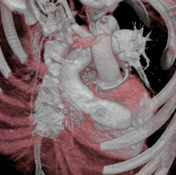
Aortic Coarctation Newborn Surgery See Image 0451203 Surgery Aortic Coarctation —  Fotos de Stock

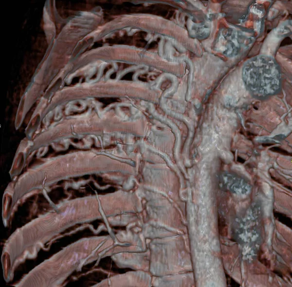
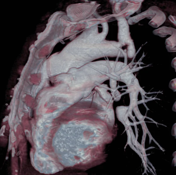
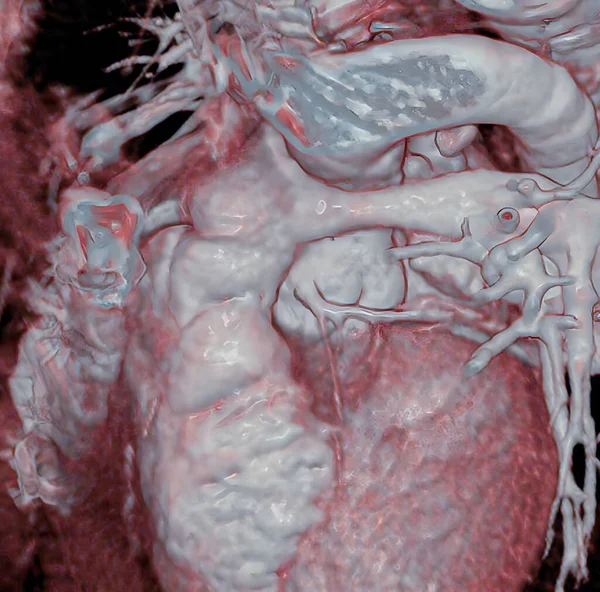
Fotos de Aortic coarctation of newborn, after surgery. See image 0451203, for before surgery. Aortic coarctation is a narrowing of the aorta, often considerable and sometimes complete, most commonly just past the point where the aorta and the subclavian arter — Imagen de stock

Aortic coarctation of newborn, after surgery. See image 0451203, for before surgery. Aortic coarctation is a narrowing of the aorta, often considerable and sometimes complete, most commonly just past the point where the aorta and the subclavian arter

 — Foto por imagepointfr

Misma serie:

Aortic coarctation is a narrowing of the aorta, often considerable and sometimes complete, most commonly just past the point where the aorta and the subclavian artery meet. Aortic coarctation causes low blood flow, often leading to arterial hypertens
Aortic Coarctation Narrowing Aorta Often Considerable Sometimes Complete Most Commonly —  Fotos de Stock
Large intercostal arteries, visible in a CT scan, developed to compensate pulmonary blood flow due to a congenital heart defect characterized by atresia of pulmonary arteries (small or absent pulmonary arteries). 3D CT scan.
Large Intercostal Arteries Visible Scan Developed Compensate Pulmonary Blood Flow —  Fotos de Stock
Renal artery stenosis (RAS) Extreme stricture (stenosis) of the left renal artery which may be the cause of severe renal-vascular arterial hypertension, 3D CT scan.
Renal Artery Stenosis Ras Extreme Stricture Stenosis Left Renal Artery —  Fotos de Stock
Pulmonary arteries and Mediastinum, heart, and aorta are visible, 3D CT scan.
Pulmonary Arteries Mediastinum Heart Aorta Visible Scan —  Fotos de Stock
Cervical vertebrae, 3D CT scan.
Cervical Vertebrae Scan —  Fotos de Stock
In the arterial switch operation (ASO), the aorta and the pulmonary artery are cut and reconnected to the \correct\ pumping chamber (ventricle). At birth the large vessels were reversed: the aorta originated in the right ventricle and the pulmonary a
Arterial Switch Operation Aso Aorta Pulmonary Artery Cut Reconnected Correct —  Fotos de Stock
3D CT scan of coronary arteries and aorta of heart.
Scan Coronary Arteries Aorta Heart —  Fotos de Stock
Thorax: Heart, aorta, and pulmonary artery are visible, 3D CT scan.
Thorax Heart Aorta Pulmonary Artery Visible Scan —  Fotos de Stock
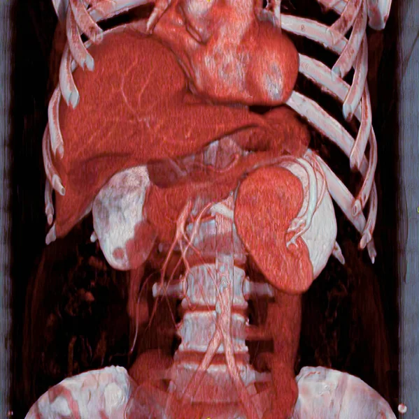
Ectopic spleen, the spleen is tipped downwards, 3D CT scan. This anomalous position is not pathological.
Ectopic Spleen Spleen Tipped Downwards Scan Anomalous Position Pathological —  Fotos de Stock
Real-time 3D reconstructions from patient images allow personalized 3D printing.
Real Time Reconstructions Patient Images Allow Personalized Printing —  Fotos de Stock
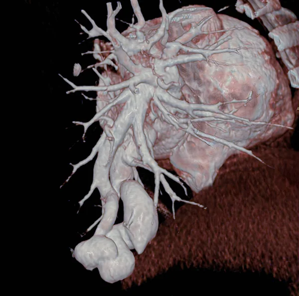
Pulmonary arteriovenous fistula is a congenital anomaly consisting of a direct communication between the pulmonary arterial and venous systems. Complications include intra-pulmonary hemorrhaging (hemoptysis). 3D CT scan.
Pulmonary Arteriovenous Fistula Congenital Anomaly Consisting Direct Communication Pulmonary Arterial —  Fotos de Stock
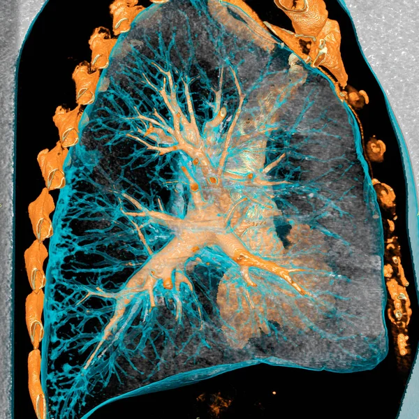
3D scanner of thoracic anatomy.
Scanner Thoracic Anatomy —  Fotos de Stock
Complex congenital cardiopathy. The 3D CT scan allows three-dimensional visualization of the heart after surgery to treat a very complicated congenital cardiopathy.
Complex Congenital Cardiopathy Scan Allows Three Dimensional Visualization Heart Surgery —  Fotos de Stock
3D CT scan of coronary bypasses.
Scan Coronary Bypasses —  Fotos de Stock
Pelvis and iliac arteries are visible, as well as a stent in the gluteal artery. 3D CT scan.
Pelvis Iliac Arteries Visible Well Stent Gluteal Artery Scan —  Fotos de Stock
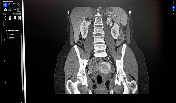
KIDNEY ABSCESS abstract background
Kidney Abscess Abstract Background —  Fotos de Stock

Información de uso

Puede usar esta foto libre de derechos "Aortic coarctation of newborn, after surgery. See image 0451203, for before surgery. Aortic coarctation is a narrowing of the aorta, often considerable and sometimes complete, most commonly just past the point where the aorta and the subclavian arter" para fines personales y comerciales de acuerdo con la Licencia Standard o Extended. La licencia Standard cubre la mayoría de los casos de uso, incluida la publicidad, los diseños de interfaz de usuario, el empaque del producto, y permite hasta 500,000 copias impresas. La Licencia Extended permite todos los casos de uso bajo la licencia Standard con derechos de impresión ilimitados y le permite usar las imágenes descargadas para mercancía, reventa de productos o distribución gratuita.

Puede comprar esta foto de stock y descargarla en alta resolución hasta 4652x4622. Fecha de importación: 11 ago 2022